
The median prescription dose was 11 Gy (10-11 Gy). Two patients had undergone surgical resection prior to gamma knife treatment. One patient with hemifacial spasm was medically controlled before gamma knife and the other two were not. All cases with trigeminal pain were receiving medication and still uncontrolled. Trigeminal pain was present in 8 patients and hemifacial spasm in 3 patients. This is a retrospective study involving 12 patients harboring cerebellopontine angle epidermoid tumors who underwent 15 sessions of gamma knife radiosurgery. The purpose of this study is to assess the safety and clinical outcome of the treatment of cerebellopontine epidermoid tumors with gamma knife radiosurgery. Radiosurgery for these tumors has rarely been reported. Intracranial epidermoid tumors are commonly found in the cerebellopontine angle where they usually present with either trigeminal neuralgia or hemifacial spasm. Gamma knife radiosurgery for cerebellopontine angle epidermoid tumors.Įl-Shehaby, Amr M N Reda, Wael A Abdel Karim, Khaled M Emad Eldin, Reem M Nabeel, Ahmed M The advantage and disadvantages of these procedures are discussed according to their usefullness in evaluating size, route of spread and localisation of cerebellopontine-angle tumors. The use of recent diagnostic procedures like scintography, CT and cisternography with oily contrast medium is critically analyzed. The value of plain films and special projections is discussed. The most important radiodiagnostic signs of cerebellopontine-angle tumors are demonstrated.

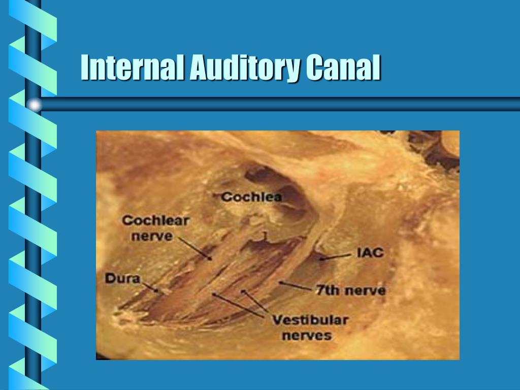
International Nuclear Information System (INIS) It remains unclear whether a non-surgical therapy such as radiation therapy is an alternative treatment option for growing choristomas.Radiodiagnosis of Cerebellopontine-angle tumors Due to a lack of access for biopsy, lesions in the IAM are often only diagnosed after surgical removal. Therefore, choristomas are usually misdiagnosed as the more common VS initially. However, choristomas cannot be distinguished from other tumors in the IAM such as VS. Osteomas are hypointense in all sequences and non-enhancing after contrast. Lipomas can usually be recognized by their hyperintense signal in T 1-weighted MRI and can be confirmed by fat suppression techniques. Vascular tumors like hemangiomas tend to be more hyperintense in MRI. Meningiomas can be distinguished from other tumors by the presence of calcification, dural tail and bone infiltration. Schwannomas of the facial nerve may extend into the labyrinthine facial nerve canal, and this feature could distinguish them from other tumors. The latter are less common in MRI at this location and include astrocytomas, ependymomas, papillomas, hemangioblastomas and metastases. Tumors in the IAM can be classified into 2 groups depending on their location: there are extra-axial lesions (outside the brain parenchyma) such as VS, meningiomas, epidermoid cysts and paragangliomas, and rare tumors such as schwannomas of the 5th, 6th, 7th, 9th, 10th, 11th or 12th cranial nerve, and vascular lesions and there are intra-axial lesions (within the brain parenchyma). How can choristomas be differentiated from other tumors in the IAM? Contrast-enhanced MRI may be helpful. Two of the patients with hearing loss presented additionally with tinnitus, 1 patient reported additionally the loss of balance and another patient tinnitus and the loss of balance.

All patients suffered from a hearing loss with the exception of 1 patient with hyperacusis and 2 additional patients in whom the hearing status was not reported. In 4 of 11 cases published in a complete clinical review, the tumors contained also skeletal muscle cells which were not found in our case. All contained smooth muscle cells as a major component, connective tissue and nerve fibers. The reports did not say whether the choristomas were removed due to growth or due to the symptoms they caused. We found only 14 reports of cases of choristomas of the 8th cranial nerve, which were all removed surgically. Smooth muscle tissue does not represent a component of cranial nerves and we suggest that the correct classification for this lesion is neuromuscular choristoma and not hamartoma. The tumor in our report consisted of smooth muscle tissue, nerve fibers and adipose tissue. Neuromuscular choristomas in the IAM are exceedingly rare lesions.


 0 kommentar(er)
